|


|
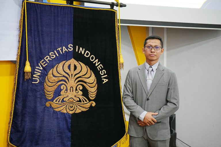
Depok, August 10, 2026 — Researchers from the Faculty of Mathematics and Natural Sciences, Universitas Indonesia (FMIPA UI), have developed a nanosystem with potential as a candidate cancer therapy by combining radiation and photothermal therapies into a single platform.
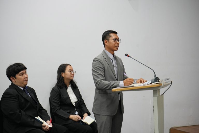
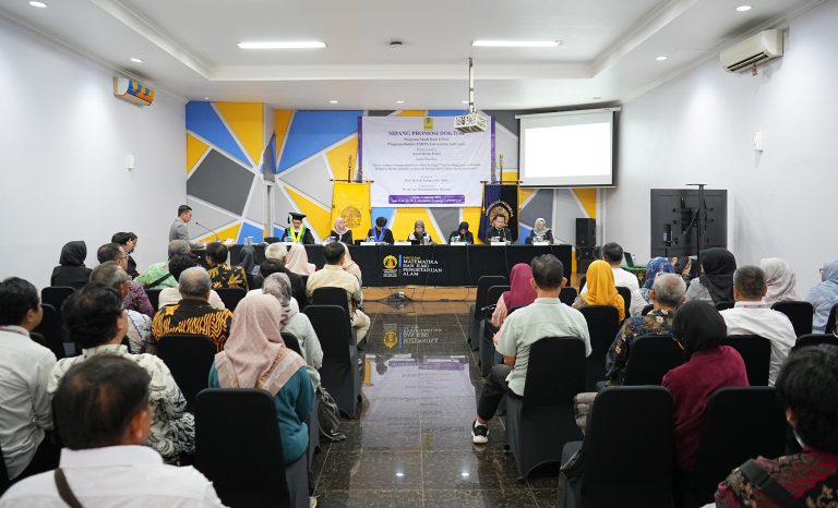
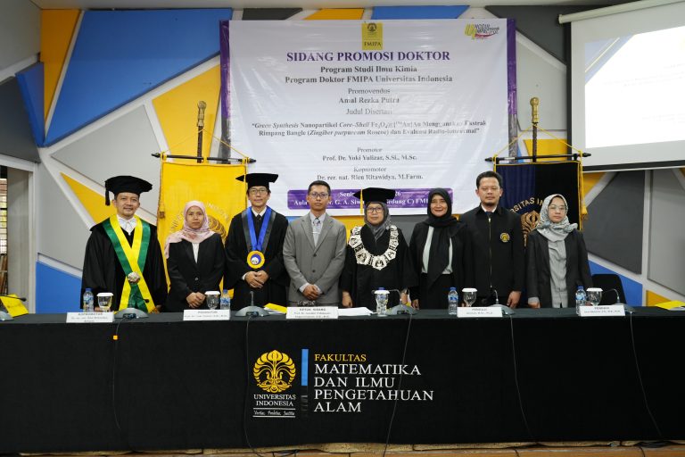
The research was developed by Amal Rezka Putra in his dissertation entitled "GREEN SYNTHESIS OF CORE-SHELL NANOPARTICLES Fe3O4@[198Au]Au USING BANGLE RHYZOME EXTRACT (Zingiber purpureum Roscoe) AND RADIO-PHOTOTHERMAL EVALUATION.” Amal defended his dissertation for the Doctoral Program in Chemistry at the Faculty of Mathematics and Natural Sciences, Universitas Indonesia (FMIPA UI), held at the Prof. G.A. Siwabessy Hall, FMIPA UI, Depok, on Monday (August 3, 2026).
In this research, Amal utilized bangle rhizome extract (Zingiber purpureum Roscoe) as a natural material to help form and stabilize nanoparticles. This approach is known as green synthesis or environmentally friendly synthesis, as it reduces reliance on synthetic chemicals that potentially pose toxic effects.
The developed nanoparticles feature a layered structure comprising an iron oxide (Fe₃O₄) magnetic core and a gold (Au) shell. This structure is then combined with radioactive gold-198 (¹⁹⁸Au) and the antibody rituximab (RTX) to form a nanosystem designed for simultaneous therapeutic and monitoring functions.
This concept is known as theranostics—an approach that combines therapeutic and diagnostic functions into a single system. With this approach, it is expected that the presence and distribution of the therapeutic agent within the body can be simultaneously monitored.
One of the advantages of the developed nanosystem is its ability to generate heat when irradiated with a laser. This heat can be utilized to destroy cancer cells in a more targeted manner through photothermal therapy.
The results of the study showed that nanoparticles with a structure core-shell Fe₃O₄@Au exhibits superior heat-generating capabilities compared to Fe₃O₄ or gold nanoparticles used individually. Upon laser irradiation at wavelengths of 532 and 980 nanometers, the nanosystem undergoes a temperature increase with a photothermal conversion efficiency of 3.2–15.5 percent.
“These findings indicate that the Fe₃O₄@Au core-shell system has the potential to be developed as a candidate photothermal agent,” concluded Amal’s research.
In addition to utilizing heat from the laser, this study incorporates the radioactive isotope gold-198 (¹⁹⁸Au). This radionuclide can deliver radiation exposure to the target area, potentially enhancing the therapeutic effect on cancer cells.
In the development of the nanosystem, Amal also tested the use of rituximab, a monoclonal antibody that can be conjugated to the surface of gold nanoparticles. In this study, rituximab was not positioned as a proven specific targeting agent for prostate cancer; rather, its use was intended to evaluate the feasibility of binding the antibody to the nanosystem's surface and to assess its stability.
The research was conducted in several stages, ranging from the synthesis of nanoparticles using bangle rhizome extract and the characterization of their physical and chemical properties to their conjugation with rituximab, the assessment of heat-generating capability, and preliminary testing on cells and the distribution of the nanosystem within the bodies of test animals.
Preliminary test results indicate that the Fe₃O₄@[¹⁹⁸Au]Au-RTX nanosystem is relatively well-tolerated by both LNCaP prostate cancer cells and normal Vero cells at low to moderate concentrations in the absence of laser irradiation.
Upon laser irradiation, the viability—or survival rate—of LNCaP cells decreased more significantly. These findings indicate that the heat generated by the nanosystem can be activated externally using a laser.
However, the study also identified a safety limit that must be considered. At a concentration of 500 micrograms per milliliter, the viability of Vero cells decreased significantly. This indicates that at high concentrations, the nanosystem begins to exert toxic effects on normal cells.
Animal testing also provides important information regarding the behavior of nanosystems within the bodies of test animals. Nanoparticles 198Au was primarily found to accumulate in the liver. Meanwhile, in systems using radioiodine tracers, a portion of the radioiodine migrated to the thyroid gland. This indicates the release of iodine from the antibody component, a process known as deiodination.
“Thus, this nanosystem holds potential as a candidate radio-photothermal agent, though it still requires optimization regarding dosage, stability, selectivity toward cancer cells, and biological safety,” the study concluded.
Amal pursued doctoral studies for seven semesters and achieved a Grade Point Average (GPA) of 4.00 with Summa Cum Laude honors.
The doctoral defense was chaired by Prof. Dr. Ivandini Tribidasari Anggraningrum, S.Si., M.Si., who also serves as the Vice Dean for Resources, Ventures, and General Administration at the Faculty of Mathematics and Natural Sciences, Universitas Indonesia (FMIPA UI). Amal was supervised by Prof. Dr. Yoki Yulizar, S.Si., M.Sc. (Department of Chemistry, FMIPA UI) as the primary supervisor (promotor) and Dr. rer. nat. Rien Ritawidya, M.Farm. (Research Center for Radioisotope, Radiopharmaceutical, and Biodosimetry Technology, National Research and Innovation Agency) as the co-supervisor.
The results of this study are still in the early stages of development. Further testing is required to determine safe and effective dosages, enhance the stability of the nanosystem, and ensure the ability to target cancer cells more selectively before advancing to testing in tumor-bearing animal models and clinical stages.